We have compiled a list of manufacturers, distributors, product information, reference prices, and rankings for Transmission electron microscope.
ipros is IPROS GMS IPROS One of the largest technical database sites in Japan that collects information on.

Transmission electron microscope - List of Manufacturers, Suppliers, Companies and Products | IPROS GMS

Transmission electron microscope Product List

1~15 item / All 15 items

Displayed results

Observation of micro-nano domain morphology using High-Resolution Transmission Electron Microscopy (HR-TEM)

We provide contract analysis services equipped with devices for physical analysis, and we also offer on-site analysis.

The main equipment includes "HR-TEM," "Q-pole type SIMS," "μESCA," and "RAMAN." We have accumulated know-how in micro and nano-level surface analysis, allowing us to provide reliable data in a short period. We also offer advice on the analysis of unknown samples. 【Observation of Micro Areas - High-Resolution Transmission Electron Microscopy (HR-TEM)】 ○ A method that irradiates a thinly processed sample with a high-speed electron beam, providing magnified images and information on crystal structures through the imaging of transmitted and scattered electrons. ○ It is suitable for evaluating the structure and crystallinity of thin films. High-resolution observation enables the examination of crystal structures. ● For more details, please download the catalog or contact us.

  • Food Testing/Analysis/Measuring Equipment
  • Transmission electron microscope

Added to bookmarks

Bookmarks list

Bookmark has been removed

Bookmarks list

You can't add any more bookmarks

By registering as a member, you can increase the number of bookmarks you can save and organize them with labels.

Free membership registration

(S)TEM (Scanning Transmission Electron Microscopy)

Elemental analysis, state evaluation, particle size analysis, and acquisition of three-dimensional structural images at the nanoscale.

TEM (Transmission Electron Microscopy) is a method that involves irradiating a thin sample with an electron beam, imaging the electrons that have passed through or scattered from the sample, and observing it at high magnification. ■ Advantages - Enlarged images can be obtained with sub-nanometer spatial resolution, allowing for the observation and analysis of the sample's fine structure and lattice defects. - It is possible to evaluate the crystallinity of the sample and identify materials. - By fabricating samples using FIB (Focused Ion Beam), it is possible to observe specific locations within a device with pinpoint accuracy. - By combining optional features, it is also possible to analyze the composition and state of localized areas. ■ Disadvantages - It is necessary to thin the sample (in some cases, thinning may be difficult for certain samples). - It does not observe individual atoms but rather displays average information in the thickness direction of the sample (typically about 0.1 μm thick). - Sample processing and observation may lead to alteration or deformation of the sample.

  • 打ち合わせ.jpg
  • セミナー.jpg
  • Contract Analysis
  • Contract measurement
  • Contract Inspection
  • Transmission electron microscope

Added to bookmarks

Bookmarks list

Bookmark has been removed

Bookmarks list

You can't add any more bookmarks

By registering as a member, you can increase the number of bookmarks you can save and organize them with labels.

Free membership registration

Types and Characteristics of Electron Diffraction

TEM: Transmission Electron Microscopy

The electron diffraction method using an electron microscope is classified into three types based on the way the electron beam is incident on the sample. The characteristics of each type and examples of data are presented. It is necessary to choose the appropriate method according to the size of the evaluation object and the analysis purpose.

  • Contract Analysis
  • Transmission electron microscope

Added to bookmarks

Bookmarks list

Bookmark has been removed

Bookmarks list

You can't add any more bookmarks

By registering as a member, you can increase the number of bookmarks you can save and organize them with labels.

Free membership registration

[Analysis Case] Polarity Evaluation of GaN by ABF-STEM Observation

Atomic-level observation is possible with the Cs collector-equipped STEM.

GaN, which is being utilized as a power device and optical device, has a hexagonal wurtzite structure and exhibits crystallographic asymmetry (Ga polarity and N polarity) in the c-axis direction. The growth processes of epitaxial films differ between Ga polarity and N polarity, and the surface physical properties and chemical reactivity of the crystal also vary. In this document, the polarity of GaN was evaluated through annular bright field (ABF)-STEM observation. As a result, the positions of the Ga sites and N sites were identified, allowing for a visual clarification of the characteristics of Ga polarity and N polarity. Measurement method: TEM Product fields: Power devices, optical devices Analysis objectives: Shape evaluation, structural evaluation, thickness evaluation For more details, please download the document or contact us.

  • Contract Analysis
  • Transmission electron microscope

Added to bookmarks

Bookmarks list

Bookmark has been removed

Bookmarks list

You can't add any more bookmarks

By registering as a member, you can increase the number of bookmarks you can save and organize them with labels.

Free membership registration

"Basics of TEM" "Applications and Case Studies of TEM" *A gift for all interested participants.

Basic knowledge and analysis examples of TEM (Transmission Electron Microscopy) used in a wide range of research and development fields such as polymers and semiconductors.

TEM (Transmission Electron Microscopy) is a method that involves irradiating a thin sample with electrons, imaging the electrons that have passed through or scattered from the sample, and observing it at high magnification. At MST, we are offering "Basics of TEM," which includes fundamental knowledge such as the characteristics of TEM and sample preparation methods, and "Applications and Examples of TEM," which features representative analysis cases and examples of combined analysis with other methods! This material is presented in an easy-to-understand manner using photographs and diagrams. [Contents (partial)] <Basics of TEM>  ■What can be understood with TEM and STEM ■Characteristics of TEM and STEM  ■Sample preparation methods ■Factors affecting contrast  ■Differentiation between TEM and STEM ■Ultra-high resolution HAADF observation <Applications and Examples of TEM>  ■Analysis examples   ・TEM analysis under controlled atmosphere and cooling   ・Evaluation of contact electrodes in SiC power MOSFETs   ・Structural evaluation of fine transistors *If you would like a booklet, please check the box for "Request for catalog" in the "Contact Us" section to apply. (The downloadable PDF material consists of the first few pages.)

  • Other microscopes
  • Transmission electron microscope

Added to bookmarks

Bookmarks list

Bookmark has been removed

Bookmarks list

You can't add any more bookmarks

By registering as a member, you can increase the number of bookmarks you can save and organize them with labels.

Free membership registration

[Analysis Case] TEM Analysis under Atmosphere Control and Cooling (B0156)

Observation with a high-resolution SEM equipped with FIB without atmospheric exposure (controlled atmosphere)!

Our organization has established a system that suppresses atmospheric exposure through atmosphere control, and further cools the samples for processing, observation, and analysis. We can produce TEM thin samples while maintaining the original structure of the specimen, allowing for observation and analysis. Even with unstable materials, we can perform thinning processing while cooling, and by maintaining a vacuum during the transfer between processing and observation devices, we enable cross-sectional TEM/SEM observation that minimizes atmospheric exposure and thermal alteration. 【Measurement and Processing Methods】 ■ [(S)TEM] Scanning Transmission Electron Microscopy ■ Processing under atmosphere control ■ Cryo-processing *For more details, please download the PDF or feel free to contact us.

  • Contract Analysis
  • Transmission electron microscope

Added to bookmarks

Bookmarks list

Bookmark has been removed

Bookmarks list

You can't add any more bookmarks

By registering as a member, you can increase the number of bookmarks you can save and organize them with labels.

Free membership registration

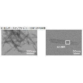
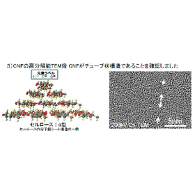
[Analysis Case] (S)TEM Observation by Negative Staining

Examples of morphological observation of liposomes, CNF, microparticles, proteins, etc.!

Our organization offers (S)TEM observation using negative staining. Organic functional materials, such as fine particles and fibrous substances, are primarily composed of light elements like C (carbon), H (hydrogen), O (oxygen), and N (nitrogen), which do not provide sufficient contrast for morphological observation in (S)TEM. Therefore, by applying staining to these materials, we can enhance the contrast and enable observation. In this instance, we will introduce a case where negative staining was performed using phosphotungstic acid (H3[P(W3O10)4]·nH2O, abbreviated as PTA). 【Measurement Method / Processing Method】 ■ [(S)TEM] Scanning Transmission Electron Microscopy ■ Others *For more details, please download the PDF or feel free to contact us.

  • 10a.png
  • 10b.png
  • Contract Analysis
  • Transmission electron microscope

Added to bookmarks

Bookmarks list

Bookmark has been removed

Bookmarks list

You can't add any more bookmarks

By registering as a member, you can increase the number of bookmarks you can save and organize them with labels.

Free membership registration

OBF STEM method (Optimal Bright Field Scanning Transmission Electron Microscopy)

OBF STEM: Optimum Bright-Field STEM Method

The OBF STEM method is a technique that reconstructs a single STEM image by applying a weighted frequency filter to multiple STEM images obtained from each segment of a segmented STEM detector, appropriately emphasizing different spatial frequency components for each segment. It is characterized by its ability to efficiently utilize the detection signal, allowing for the visualization of structures with low noise and high contrast even under conditions of low electron beam exposure.

  • img_B0310_7.jpg
  • Contract Analysis
  • Secondary Cells/Batteries
  • Memory
  • Transmission electron microscope

Added to bookmarks

Bookmarks list

Bookmark has been removed

Bookmarks list

You can't add any more bookmarks

By registering as a member, you can increase the number of bookmarks you can save and organize them with labels.

Free membership registration

Spherical aberration correction function

TEM: Transmission Electron Microscopy

In a STEM device equipped with a spherical aberration correction function (Cs corrector), high-resolution observation and high-sensitivity analysis at the atomic level are possible. The resolution is approximately 0.10 nm.

  • Contract Analysis
  • Transmission electron microscope

Added to bookmarks

Bookmarks list

Bookmark has been removed

Bookmarks list

You can't add any more bookmarks

By registering as a member, you can increase the number of bookmarks you can save and organize them with labels.

Free membership registration

[Analysis Case] STEM/EDX and Image Simulation for Crystal Structure Evaluation

The evaluation of the crystal structure can be performed based on the STEM images and the results of atomic composition measurements.

By measuring the sample, it is possible to evaluate the crystal structure through the combination of results obtained and simulations. This document introduces a case study in which the crystal structure is discussed by comparing the results obtained from HAADF-STEM and EDX measurements on polycrystalline neodymium magnets with simulated images using the respective measurement conditions. The combination of measurement results and computational simulation results allows for a deeper understanding of the crystal structure.

  • Contract Analysis
  • magnet
  • Memory
  • Transmission electron microscope

Added to bookmarks

Bookmarks list

Bookmark has been removed

Bookmarks list

You can't add any more bookmarks

By registering as a member, you can increase the number of bookmarks you can save and organize them with labels.

Free membership registration

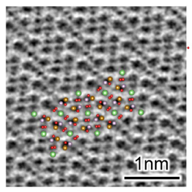
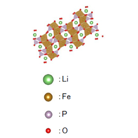
[Analysis Case] Local Area Evaluation of Positive Electrode Active Material in Secondary Batteries (C0614)

Composition, crystal structure, and chemical state evaluation near the surface of living materials are possible!

Our company conducts local area evaluation of positive electrode active materials for secondary batteries (C0614). It is known that in lithium-ion secondary batteries, the crystal structure of the active material surface and the valence of metal elements change due to ion extraction and insertion during charge and discharge, as well as degradation. In a case where TEM was used for local area evaluation to assess composition, crystal structure, and chemical state, it was found through EDX that AlOx is coated on the outermost layer. Additionally, electron diffraction and EELS revealed that the crystal structure and valence differ between the surface and the interior of the active material. [Measurement and Processing Methods] ■ [(S)TEM] Scanning Transmission Electron Microscopy ■ [TEM-EDX] Energy Dispersive X-ray Spectroscopy (TEM) ■ [TEM-EELS] Electron Energy Loss Spectroscopy *For more details, please download the PDF or feel free to contact us.

  • Contract Analysis
  • Transmission electron microscope

Added to bookmarks

Bookmarks list

Bookmark has been removed

Bookmarks list

You can't add any more bookmarks

By registering as a member, you can increase the number of bookmarks you can save and organize them with labels.

Free membership registration

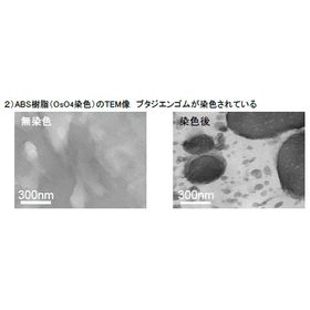
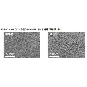
[Analysis Case] (S)TEM Observation of Polymer Materials Using Electronic Staining

We will introduce examples of observing polymer materials using SEM-STEM and (S)TEM!

Our organization offers (S)TEM observation of polymer materials using electron staining. Since polymer materials are composed of light elements, it is difficult to obtain clear contrast in (S)TEM observations. Staining can enhance the contrast for such materials. This document introduces examples of staining and observation using a method called electron staining with ruthenium tetroxide (RuO4), osmium tetroxide (OsO4), and phosphotungstic acid (H3[P(W3O10)4]·xH2O, abbreviated as PTA). [Measurement and Processing Methods] ■ [(S)TEM] (Scanning) Transmission Electron Microscopy ■ Others *For more details, please download the PDF or feel free to contact us.

  • 9a.png
  • 9b.png
  • Contract Analysis
  • Transmission electron microscope

Added to bookmarks

Bookmarks list

Bookmark has been removed

Bookmarks list

You can't add any more bookmarks

By registering as a member, you can increase the number of bookmarks you can save and organize them with labels.

Free membership registration

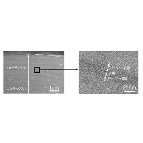
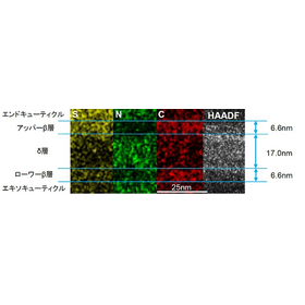
[Analysis Case] Fine Structure of Hair Cross-Section (S)TEM Analysis

We will introduce examples of observations of hair cell membrane complexes (CMC)!

Our organization offers (S)TEM analysis of the fine structure of hair cross-sections. The hair cell membrane complex (CMC) serves as a pathway for chemicals during processes such as bleaching and dyeing hair. In particular, the CMC between the cuticle layers has a three-layer structure, which we have visualized. We present a case where osmium tetroxide (OsO4) was used for electron staining targeting unsaturated fatty acids in lipids, followed by TEM observation and STEM-EDX analysis. [Measurement and Processing Methods] ■ [(S)TEM] Scanning Transmission Electron Microscopy ■ [TEM-EDX] Energy Dispersive X-ray Spectroscopy (TEM) ■ Ultramicrotome processing ■ Others *For more details, please download the PDF or feel free to contact us.

  • 11a.png
  • 11b.png
  • Contract Analysis
  • Transmission electron microscope

Added to bookmarks

Bookmarks list

Bookmark has been removed

Bookmarks list

You can't add any more bookmarks

By registering as a member, you can increase the number of bookmarks you can save and organize them with labels.

Free membership registration

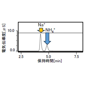
[Analysis Case] Evaluation of Solution Components Before and After Adsorption and Zeolite Structure

We capture the ion exchange process from the ion content in the solution and the structural changes of the adsorbent.

Zeolites are porous crystalline aluminosilicates with fine pores of a few nanometers, widely used as adsorbents for molecular adsorption in fine pores and cation exchange within the crystal for water treatment. Due to the freedom of design, they can be tailored to specific purposes; however, evaluating whether the performance aligns with the design requires an assessment from both a macroscopic perspective of water components and a microscopic perspective of the adsorbent's fine structure. This analysis presents a case study of an immersion experiment of zeolite in ammonia water, capturing the changes in ion content in the water before and after immersion, as well as the structural changes in the zeolite.

  • img_C0745_2.jpg
  • Contract measurement
  • Structural Analysis
  • Evaluation Board
  • Transmission electron microscope

Added to bookmarks

Bookmarks list

Bookmark has been removed

Bookmarks list

You can't add any more bookmarks

By registering as a member, you can increase the number of bookmarks you can save and organize them with labels.

Free membership registration

Search Keywords Related to Transmission electron microscope